


Pressed Ceramic Veneer and Microscope-Assisted Root Canal Treatment


What brought the patient to our clinic?
Our patient’s dental problems were caused by their sporting activities. Repeated frontal impacts resulted in a fractured tooth. The patient sought an aesthetic solution to this damage.
Diagnosis and Proposed Treatment
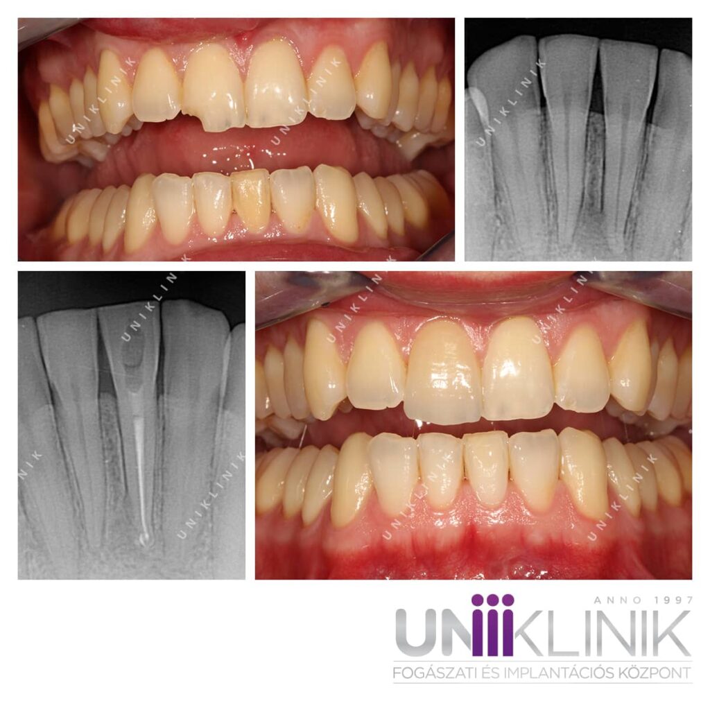
The effects of repeated frontal trauma were clearly visible on the front teeth. A significant portion of the upper right central incisor’s crown had broken off. Additionally, one of the lower incisors appeared several shades darker than the surrounding teeth—an indication of pulp necrosis (a dead tooth).
Radiographic imaging confirmed the diagnosis: a dark area around the apex of the lower left central incisor pointed to inflammation.
How did the procedure go?
The first step was to eliminate the inflammation through microscope-assisted root canal treatment. Success was confirmed by a follow-up X-ray, which showed that the dark area had resolved, indicating healing.
To address the discoloration, internal tooth whitening was performed. In this process, a whitening agent is placed inside the cavity created during the root canal, then sealed with a temporary filling. This technique is safe, as the bleaching agent remains entirely within the tooth. The material was replaced every two weeks until the desired shade was achieved—two applications in this case.
To restore the broken upper incisor, a pressed ceramic veneer was chosen. In addition to its excellent aesthetic properties, this material is durable and well-suited to withstand the mechanical stresses likely to recur in the patient’s active lifestyle.
To prevent future trauma, we also fabricated a custom sports mouthguard in our dental laboratory. This helps protect not only the new veneer but also the remaining natural teeth—a top priority in our care philosophy.

Both professionally and personally, the care was high-quality, kind, and quick.
Bálint (36)
Excellent technical equipment and a wide range of services available quickly and conveniently on site.
Béla (45)