


Dental CT: X-Ray and CBCT Imaging, Digital Diagnostics
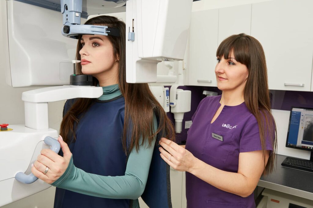
Our clinic is equipped with a state-of-the-art radiology center. High-resolution imaging is essential for accurate dental treatment as it allows for precise diagnosis and provides crucial information for your dentist. We prioritise the use of advanced technology to ensure that all imaging is performed safely and efficiently.
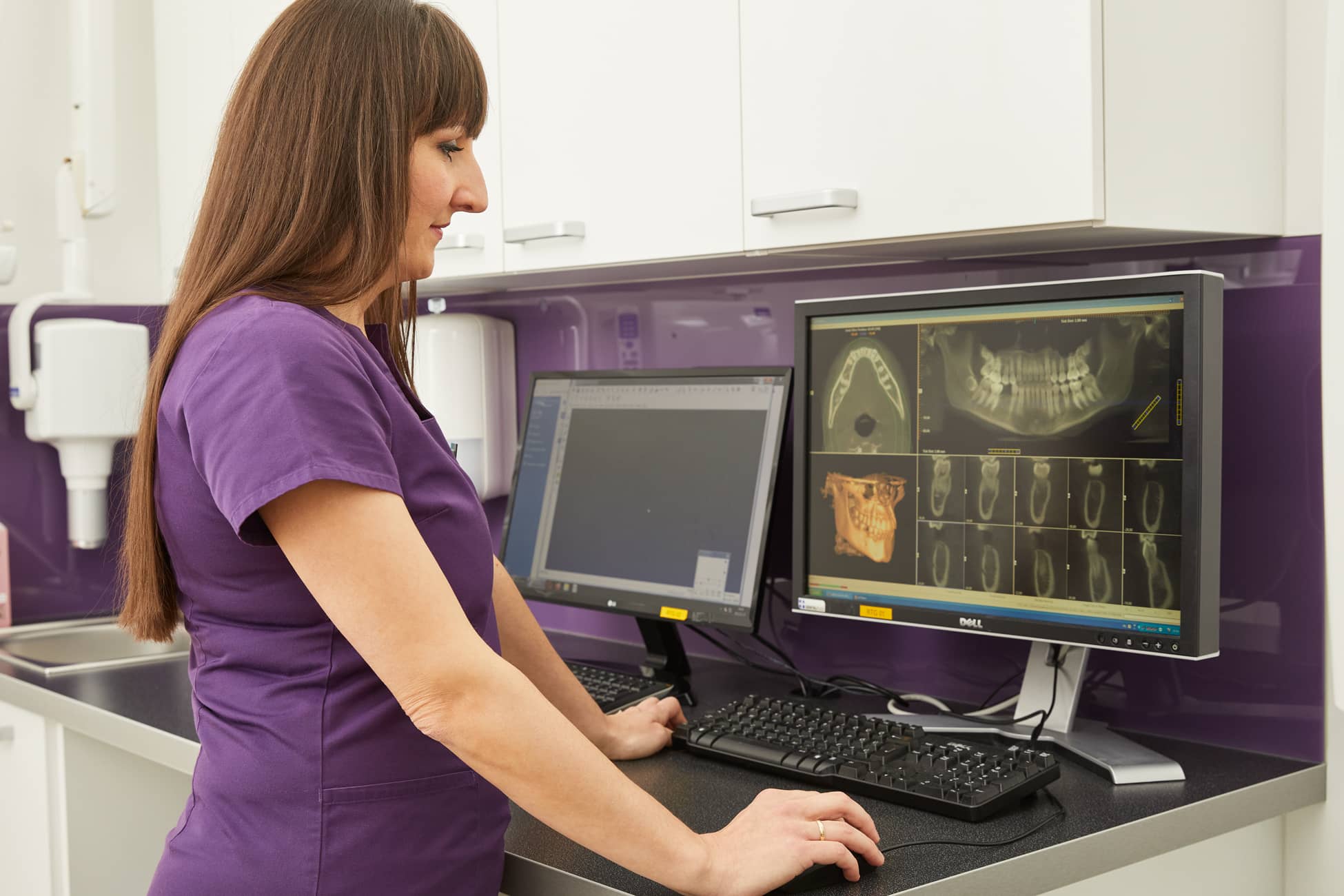
DEXIS OP 3D LX: the next generation of cone beam technology
Our radiology department is equipped with the latest DEXIS (formerly KaVo) OP 3D LX system, which utilises AI technology for imaging. Its innovative SMARTVIEW function simplifies image capture, enhancing efficiency and patient care.
What is CBCT?
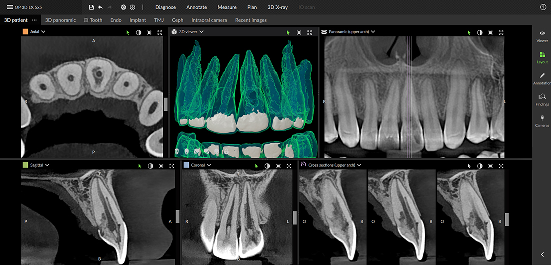
Cone Beam CT (CBCT) stands for cone beam computed tomography, a non-invasive imaging technique used in dentistry. During the procedure, the CT scanner emits a cone-shaped X-ray beam, capturing a series of two-dimensional images from various angles around the patient’s head. These images are then reconstructed to create a three-dimensional view of the oral and maxillofacial structures, including teeth, bone, nerves, and soft tissues. This comprehensive visualization allows for more accurate diagnosis and precise treatment planning.
Traditional panoramic X-rays can produce image distortions, making evaluation more challenging. In contrast, a dental CT scanner provides a proportionally accurate, distortion-free, three-dimensional image that can be viewed from multiple angles. It also reveals not only the quantity but also the quality of the bone. Additionally, CBCT exposes patients to lower radiation levels, as these devices do not emit radiation continuously like conventional dental CT scanners. Instead, CBCT operates with minimal radiation dose, ensuring a safer imaging process.
What are the advantages of CBCT over traditional two-dimensional panoramic X-rays?
Traditional panoramic X-rays distort different areas of the image to varying degrees, depending on the equipment used. Additionally, all anatomical structures positioned between the detector and the X-ray source are superimposed onto the image. This can obscure certain details or make them appear blurry and undefined. These overlapping structures significantly complicate the interpretation of the radiograph.
This advanced imaging technique allows for a true-to-scale (1:1) 3D representation of the examined area without superimposition, viewable from multiple angles. Additionally, it provides valuable information not only about bone quantity but also about bone quality.
Why is CBCT better than medical CT?
CBCT imaging exposes the patient to minimal radiation compared to conventional medical CT scans, which are used for examining various body regions such as the spine or skull. The radiation dose from CBCT is equivalent to that of standard dental imaging techniques and is only about 1% of the dose from medical CT. Additionally, while a medical CT scan takes several minutes, CBCT scanning is completed in just 20–40 seconds.
How is a CBCT scan performed?
The CBCT device consists of a computer, an X-ray source, and a detector positioned opposite it. Depending on the settings, it performs a 360˚ rotation around the patient’s head in 10-40 seconds, similar to the function of panoramic X-ray machines. In contrast to traditional CT scanners, where imaging occurs layer by layer during the 360˚ rotation, the CBCT uses a cone-shaped X-ray beam to create a series of two-dimensional images, from which it constructs a three-dimensional database. It does not expose the patient continuously like traditional CT scanners; instead, it captures images at specific angles. This results in a significantly lower radiation dose to the patient, which is of great importance.
The DEXIS OP 3D LX dental CT machine: a new level in dental imaging
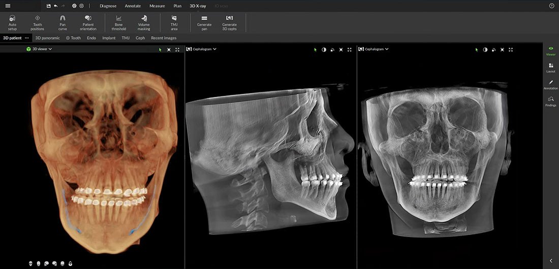
This 3D technology-based multimodal imaging platform covers the full spectrum of dental imaging needs, from endodontics to the most complex implant cases.
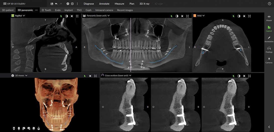
This next-generation system offers a flexible field of view (FOV), allowing for a variety of scans: from partial scans of 3-4 teeth (5 x 5 cm) to a full-face CBCT scan (15 x 20 cm), which currently represents the largest field of view available with the DEXIS OP 3D.
The 15×20 cm full-face scan is taken in a single exposure, not by stitching two images together, resulting in a lower radiation dose.
Get to know the cutting-edge DEXIS OP 3D LX
3D imaging options with the DEXIS OP 3D LX
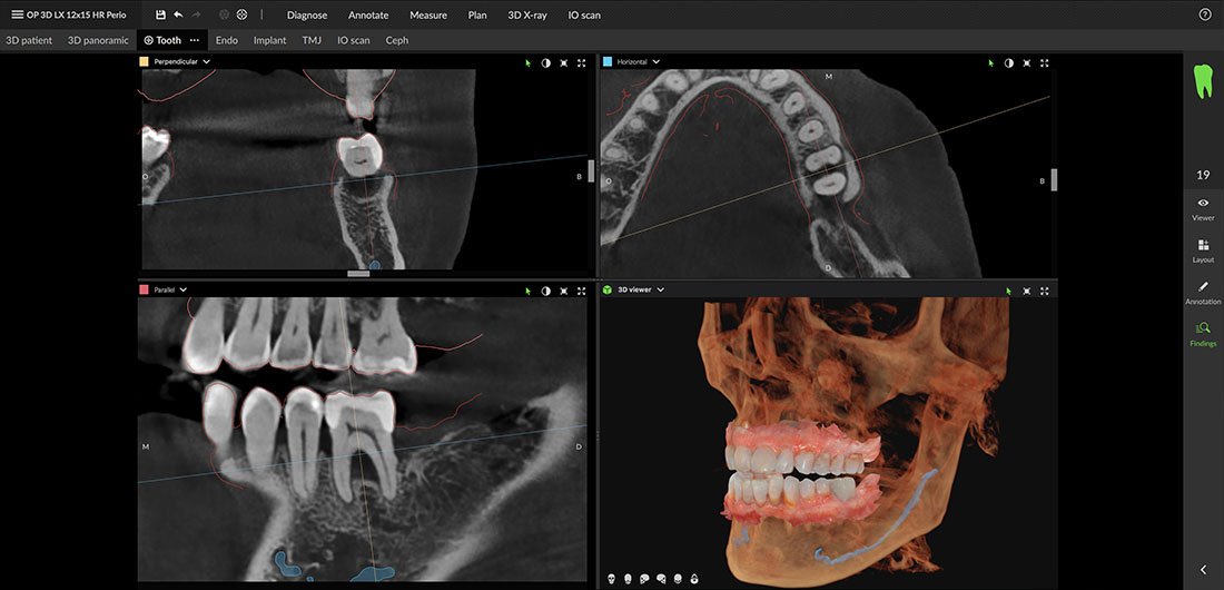
Endodontics
With its dedicated endodontic segmentation and precise scanning positioning, the DEXIS OP 3D LX is optimized to display even the smallest details, which can be crucial for endodontic diagnosis and treatment.
Maxillofacial surgery
Before complex surgical procedures, clear and precise data helps determine bone quality and size, as well as anatomical structures, allowing the surgery to be planned and executed with the highest accuracy.
Orthodontics
The evaluation of orthodontic treatments is clearer with high-resolution CBCT imaging. The DEXIS OP 3D LX aids in diagnosing complex cases by providing the most accurate positioning of unerupted or impacted teeth.
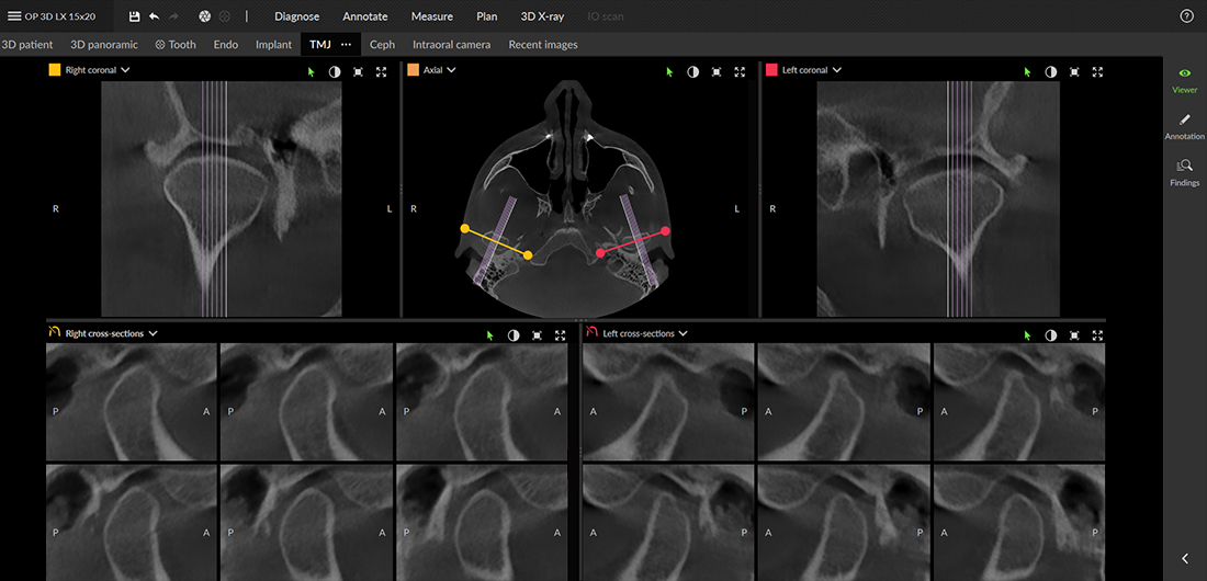
Gnathology
The device is also suitable for the bilateral visualization of the temporomandibular joint, assessing degenerative changes in the hard tissues, and evaluating occlusion positioning. These all support gnathological, or temporomandibular joint, diagnosis and treatment.
Periodontology
The OP 3D LX allows for a comprehensive examination of the bone structure, as well as the positioning of the sinus and nerves, ensuring accurate determination of treatment options for bone loss.
Prosthodontics
The system supports perfect visualization by enabling the specialist to integrate 3D data with intraoral scan information, providing a complete view of the patient’s anatomy.
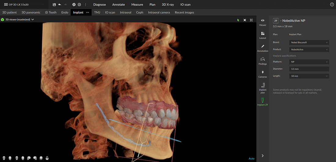
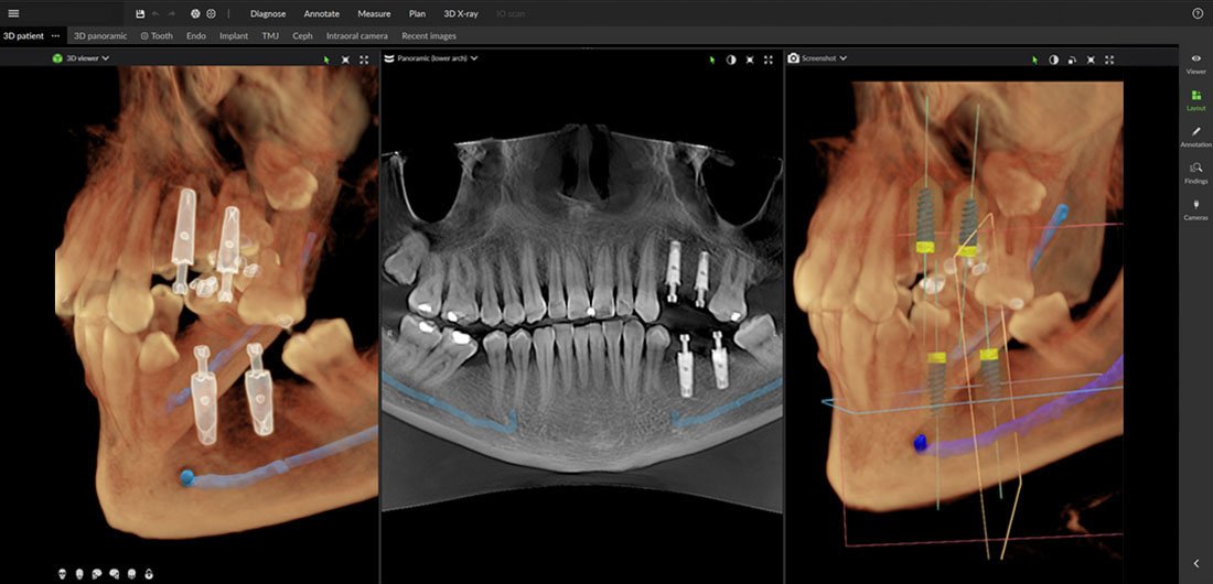
Implant placement
Whether it’s a single implant placement or the application of the All-on-4 technology, the DEXIS OP 3D LX enables multidimensional imaging for pre-surgical diagnosis and supports the most precise implant placement.
The steps of performing a dental CT scan
-
- Preparation
The patient must remove all metal objects (jewelry, glasses, removable dental prostheses) from the head and neck area to avoid interference with the scan. - Positioning
The patient sits or stands in a special chair, wearing a lead apron, while the assistant stabilizes the jaw for the scan. - Imaging
The CT machine performs a 360-degree rotational movement around the patient’s head, emitting X-rays and recording the reflected radiation with detectors, a process lasting 20-40 seconds. - Data processing
The machine converts the collected data into a three-dimensional image using computer software. - Analysis of results
The dentist or radiologist analyzes the resulting images and uses them to create a diagnosis or treatment plan.
- Preparation
Dental CT prices
3D CT felvétel
Ez a képalkotó eljárás a ma elérhető legpontosabb három dimenziós felvételt adja a fogazatról, állcsontokról, arcüregről. Ennek a rendkívül precíz vizsgálatnak az eredményeként a fogászati szakemberek teljesen pontos képet kapnak a beavatkozás helyéről, így lehetőségük van annak minden lépését előre megtervezni. Segítségre van szájsebészeti, endodonciai, fogszabályzási esetekben, illetve góckutatáskor is.
Részleges CBCT felvétel (3-4 fog) 5×5
22 500 FT
Felső állcsont CBCT felvétel (maxilla) 6×9
24 500 FT
Alsó állcsont CBCT felvétel (mandibula) 6×9
24 500 FT
Kompakt fogsor CBCT felvétel (Front és kisőrlő fogak területéről) 8×8
26 500 FT
Kis térfogatú teljes fogászati CBCT felvétel 10×10
29 500 FT
Nagy térfogatú standard CBCT felvétel 12×15
32 500 FT
Teljes arckoponya CBCT felvétel (Maxillofacialis komplex) 15×20
35 500 FT
CBCT felvétel implantációs sablonról is
+5 500 Ft / állcsontonként
Röntgen felvételek, digitális diagnosztika
A diagnózis felállításához, a kezelési tervhez vagy a páciens aktuális állapotának ellenőrzéséhez különböző röntgenfelvételek szükségesek a fogakról és állcsontokról. RVG, panorámaröntgen, 3D-CT – ez csak néhány a speciális felvételek közül, amelyeket saját laborunkban készítünk el.
Panoráma röntgen (OP-felvétel)
10 000 Ft
Periapicalis felvétel (kisfog felvétel)
2-3 fog felvétele esetén; egyszerre készített felvétel; filmtartóval készül; higiénikus; ülő helyzetben végzett; pár percet vesz igénybe.
4 500 Ft
Carieskereső felvétel
Caries keresésére, alsó-felső fogak koronáiról; kontaktfelszín felvétele; ülő helyzetben végzett; 6-7 percet vesz igénybe.
4 500 Ft
Teljes státusz felvétel
Több kis felvétel készül, amelyek célja, hogy minden fog látható legyen a felvételeken; higiénikus; ülő helyzetben végzett; 10-12 percet vesz igénybe.
50 000 Ft
Sinus felvétel
Az arcüreg és orrmelléküreg röntgenfelvétele; álló helyzetben végzett; 5-6 percet vesz igénybe.
10 000 Ft
TMI felvétel
Az állkapocsízület felvétele; álló helyzetben végzett; 5-6 percet vesz igénybe.
10 000 Ft
Teleröntgen felvétel
Koponyafelvételről van szó, amelyet elsősorban fogszabályzáshoz kérnek; oldalirányú; álló helyzetben végzett; 5-6 percet vesz igénybe.
10 000 Ft
Postero-anterior felvétel
Szemközti koponyafelvétel; álló helyzetben végzett; 5-6 percet vesz igénybe.
10 000 Ft
Digitális felvétel kiadása a fogászat által biztosított pendrive-on
3500 FT
The most cutting-edge dental CT in Budapest, at Uniklinik
Our radiology center uses the state-of-the-art DEXIS OP 3D LX system, which incorporates AI technology for capturing images. Its innovative SMARTVIEW feature simplifies and enhances image acquisition, making the process more efficient.
Contact us by phone or request a consultation via our booking form so we can provide you with the best and most professional care. At our practice, we offer the latest treatments in aesthetic dentistry, from smile design to tartar removal.
Our dental clinic is equipped with its own X-ray and CT lab, allowing us to perform all necessary imaging for precise diagnosis, ranging from simple X-rays to panoramic X-rays and 3D CT scans.